EuroSafe Imaging at ECR 2017 – review
ECR 2017 showcased EuroSafe Imaging’s achievements and way forward
This year’s European Congress of Radiology was again a stunning congress and in particular a great success for EuroSafe Imaging in raising awareness of radiation protection among congress attendees. ECR 2017 clearly showed that EuroSafe Imaging’s activities help move issues related to quality and safety in medical imaging higher on people’s agenda, making radiation protection more and more relevant in the daily practice of all imaging professionals. Building on these achievements, EuroSafe Imaging is gaining even more momentum with a lot of new endeavours ahead.
EuroSafe Imaging enjoyed a great presence at ECR 2017, with five dedicated and several related sessions, as well as the electronic poster exhibition and oral presentations to promote radiation protection and safety in medical imaging among congress attendees. The highlight of ECR 2017 was the new EuroSafe Imaging Café, which was a very popular spot for networking and learning about the ESR’s quality and safety initiative.
In the following, you will find a detailed report about EuroSafe Imaging’s activities at ECR 2017.
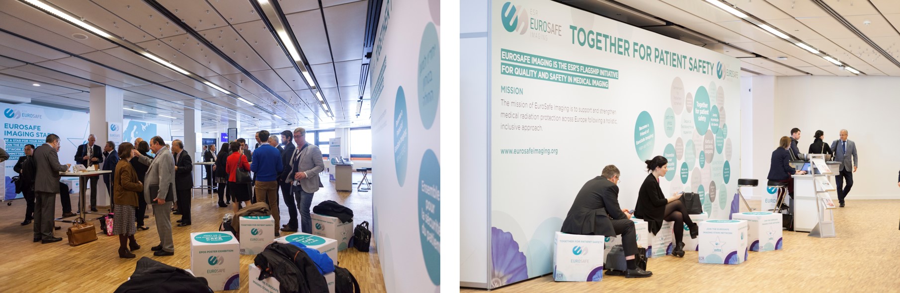
EuroSafe Imaging Café
For the first time, the ECR featured the so-called ‚EuroSafe Imaging Café‘. This café, open to all congress attendees, was not simply a place for enjoying a coffee during the breaks, but a dedicated meeting place for anyone interested in learning about radiation protection in general and EuroSafe Imaging’s activities in particular. The café also featured an information booth with plenty of material about the initiative, as well as computer terminals to browse through the electronic EuroSafe Imaging poster exhibition. The café was a very popular and well frequented spot, which contributed to raising awareness among congress attendees and thus promoting radiation protection in medical imaging.
In addition, dedicated networking events organised in the café allowed for informal discussions and exchange of experiences and ideas among EuroSafe Imaging stakeholders and contributors.
EuroSafe Imaging poster exhibition
Over 60 posters from submitters in Europe and across the world were displayed at the ECR 2017 EuroSafe Imaging Poster Exhibition, using the ESR’s Electronic Presentation Online System (EPOS). ECR 2017 also featured a Voice of EPOS session, giving selected authors the chance to present their poster during ECR.
Browse through the ECR 2017 EuroSafe Imaging posters here or watch the recording of the oral poster presentations here.
EuroSafe Imaging sessions
In 2017, five scientific sessions were categorised as EuroSafe Imaging sessions, while a number of related sessions were also promoted as part of the campaign. These sessions included renowned speakers and moderators in the field of radiation protection presenting the wide scope of EuroSafe Imaging with a particular focus on the activities carried out since ECR 2016. All sessions were very well received by the attendees, who participated in lively discussions at the end of each session.
Click here for the full ECR 2017 programme, and if you want to listen to the recorded sessions visit ECR Online (Access is free of charge, but registration is required).
![]()
Please find below short reports on the dedicated EuroSafe Imaging sessions:
1. Clinical Diagnostic Reference Levels (recording): With the new Basic Safety Standards Directive (Council Directive 2013/59/Euratom), Diagnostic Reference Levels (DRLs), which are an important tool for optimisation, have been included in European legislation. Today, most DRLs are based on modalities and anatomical body regions. In the beginning, this session gave an overview of different definitions for clinical/local/national/regional/European/global DRLs. Thereafter, the importance of the establishment of clinical DRLs, which take into account the different clinical tasks of the same area, was pointed out. Setting up clinical DRLs is more complex, as in addition to image quality the complexity and duration of a procedure have a major impact. In addition, the classification of patient conditions is required as different dose levels for one anatomical region are reasonable. Such a process will increase the number of DRLs but probably reduce the overall dose to patients. Currently, there is still a lack of concrete indication-based DRLs. The ESR has performed a pilot survey to overcome this deficit, concentrating on CT examinations of a few frequent indications. The preliminary results of the survey were presented to the participants. The session concluded with a presentation on the American College of Radiology’s work to determine DRLs and achievable doses for the 10 most common adult CT examinations in the US with the ACR Dose Index Registry data.
2. Focus on appropriate image quality: what we have to know (recording): Defining the appropriate image quality in diagnostic imaging (e.g. CT) is a very challenging task, as many factors, such as the diagnostic task, patient size and technical parameters, can impact the image quality. This session provided an insight into appropriate image quality and gave a physicist’s and radiologist’s perspective. It was pointed out that image quality should be measured as objectively as possible to be able to a) compare systems, b) assure consistent quality, c) have direct access to procedure optimisation outcomes. The measured image quality should be directly related to the diagnostic quality. Different metrics and methods, such as Fourier-based and task-based image quality metrics, are available, although they have different advantages and limitations. It was noted that the goal should be to choose an image quality that is good enough for the diagnostic task at hand, at a radiation exposure that is as low as reasonably achievable according to the ALARA concept. One way to do this is to adjust equipment settings such as tube current (mA) or tube potential (kV). Another approach is to improve the diagnostic potential of a method without necessarily reducing the dose. The session was concluded with a presentation of the review process of the Antwerp University Hospital, which includes the assessment of image quality. The session made clear that it is difficult to define appropriate image quality and „optimal“ dose due to a variety of influencing factors, and that collaboration between radiologists and physicists is essential.
3. European Alliance for Medical Radiation Protection Research (EURAMED) (recording): This session introduced EURAMED, a joint initiative of EIBIR and European associations involved in the application of ionising radiation in medicine, launched in September 2016. EURAMED is a consortium consisting of the European Association of Nuclear Medicine (EANM), the European Federation of Organisations for Medical Physics (EFOMP), the European Federation of Radiographer Societies (EFRS), the European Society of Radiology (ESR) and the European Society for Radiotherapy and Oncology (ESTRO) with the goal of jointly improving medical care and radiation protection issues through sustainable research efforts. The initial major step to overcome the fragmentation and lacking visibility of radiation protection in the medical field was the development of a first edition of a strategic research agenda (SRA) for medical radiation protection. In the course of this session, some specific topics and tasks were exemplarily presented focusing on circulating biomarkers reflecting dose exposure, general physical principles used for optimisation and dose distribution in interventional radiology.
4. European CT Dose Repository (recording): A EuroSafe Imaging working group on the development of a European CT dose repository has been established to analyse tools for automatic dose monitoring and the most frequent pitfalls. A further aim is to provide recommendations to assure reliability of statistics obtained from dose monitoring systems. In the medical area, technological and scientific developments lead to an increase in overall population exposure. Thus, the need to implement Radiation-Dose-Index Monitoring (RDIM) systems for the most important ionising-radiation procedures in connection with stochastic and deterministic risks, has become important. These software systems store Radiation-Dose-Index (RDI) data from ionising radiation modalities in a database along with patient demographic and exam information allowing the final user to visualise the RDI according to study type and patient. These data can be used both for quality assurance procedures in the diagnostic department and as benchmarks in the regional and national registries. This session highlighted that dose tracking tools are relatively new and that practical impacts vary. It was noted that these tools help to comply with legislation and are an efficient method of standardising radiation safety procedures. It was also pointed out that the responsibilities of radiographers and radiologists basically remain the same, but dose tracking can assist optimisation and dose awareness. The session concluded with a presentation on the American College of Radiology’s Dose Index Registry (DIR), which was launched in 2011 as a tool for quality improvement, so facilities can review dose indices and optimise protocols. The key features of the DIR is the automatic extraction of dose indices, patient features (e.g. gender, age, exam type, etc.) and technical parameters.
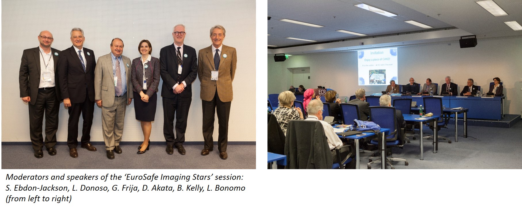
5. EuroSafe Imaging Stars (recording): The EuroSafe Imaging Stars concept was launched in the beginning of 2016. This session gave an overview of the progress and activities since its launch. In doing so, also limitations and possibilities for improvement were examined. The session highlighted the value of achieving Star status, e.g. revisiting the principles for radiation protection to perform safe imaging, self-auditing and identifying weaknesses, awareness of and commitment to radiation protection, etc. All facilities participating in the initiative are requested to complete the ‚Is your Imaging EuroSafe?‘ surveys to assess the status quo in CT practice in Europe and to build a European repository based on dose exposures for specific clinical indications to be used for benchmarking. Preliminary results of the surveys are already available. It was noted that the surveys are a good pilot project. However, the results have some limitations, e.g. high heterogeneity of data and small data sample. Also, possibilities for improvement of the Stars concept were presented, e.g. the expansion of the concept to specialised facilities (e.g. paediatric or orthopaedic hospitals), different supporting activities for the Stars network, and contributions to ESR and EuroSafe Imaging projects. Also, it was indicated that the current self-assessment to become a Star facility can be changed to an audited process. In this context the audit pack developed by the ESR Audit & Standards subcommittee to help facilities get started and to demonstrate how easy audit can be was presented. It is envisaged to pilot the audit pack within the network of Star facilities. The session concluded with a presentation on the integration of radiation protection in the clinical setting. It was pointed out that now the paradigm is changing, i.e. from producing to using guidelines. Therefore, the use of Clinical Decision Support is essential. For one anatomical location, e.g. chest CT, various different clinical indications, protocols and exposures are possible. Thus, it was emphasised that exposure protocols should be primarily based on clinical indications and not on anatomical location, and consequently, DRLs should be also established for clinical indications. Moreover, it was noted that besides national DRLs, also local DRLs (incl. local dose repository) should be established as a quality management tool, and the differences between national and local DRLs were explained. The talk concluded with a summary of the clinical components of radiation protection: Justification, image quality and risks and benefits are clinically driven; the clinical indication drives the protocol; modern tools for dose recording provide insights in the daily clinical practice.